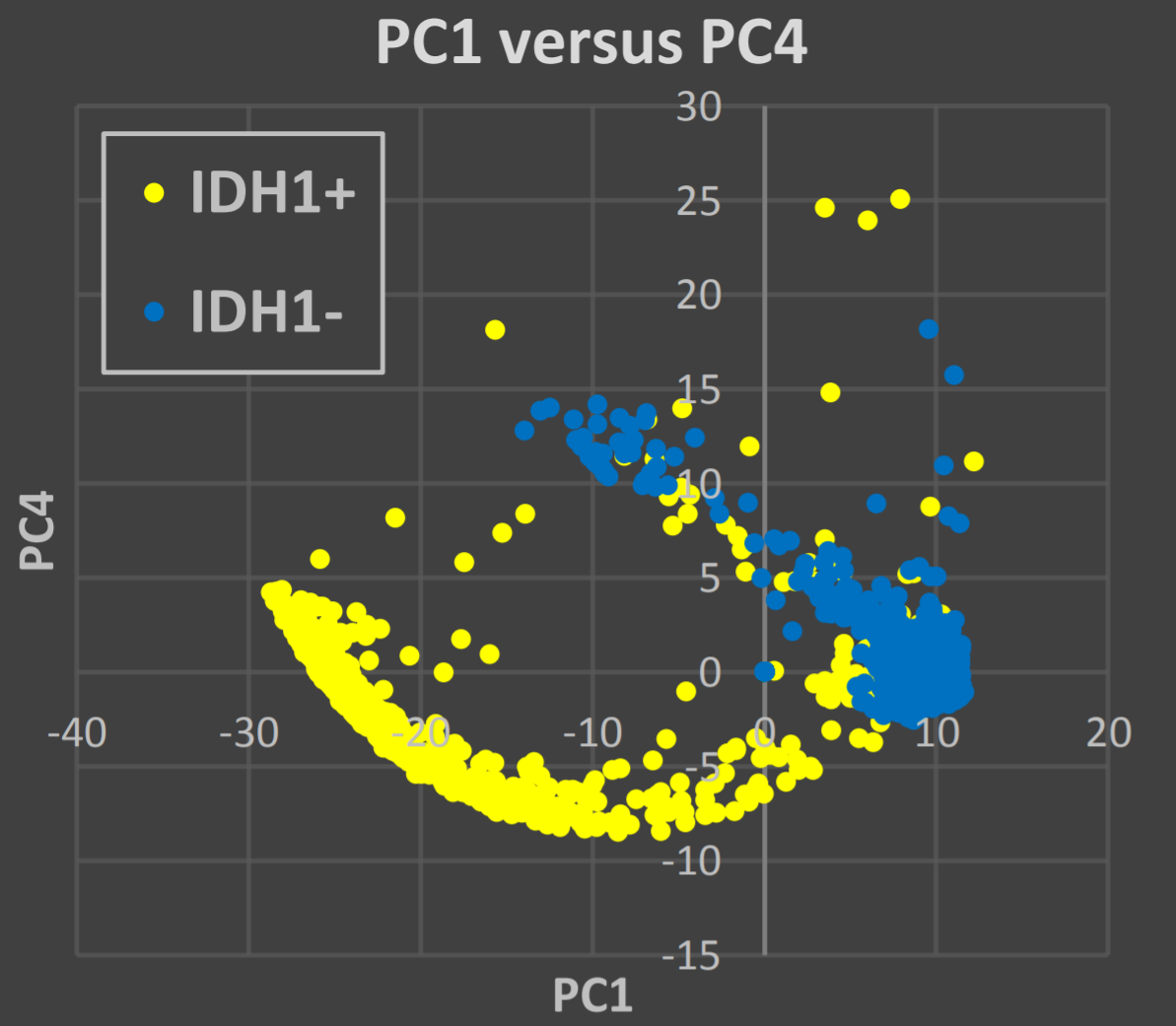
Raman spectroscopy for the identification of isocitrate dehydrogenase (IDH) mutation in glioblastomas.
 Image credit: From Abstract
Glioblastoma, a highly malignant primary brain tumor, can be stratified by the presence or absence of mutations in isocitrate dehydrogenase (IDH) 1 or 2. Establishing the presence of IDH mutations has become essential in providing accurate histopathological tumor diagnosis. Determination of IDH status is critical for clinical decision-making with evidence that the presence of IDH mutation confers better prognosis and a better response to chemotherapy1 . Conventional methods for IDH mutation detection, including immunohistochemistry and DNA sequencing, are time- and labor-consuming. In this study, we investigate the feasibility of using Raman spectroscopy to differentiate between IDH1 positive (IDH1+ ) and negative (IDH1- ) tumors through classification modelling. Forty biopsies of glioblastoma (WHO IV) in patients under the age of 40 years were classified into either IDH-1(R132H) positive or negative using standard immunohistochemistry. 6µm thick sections were prepared from formalin fixed paraffin embedded specimens. Based on the hematoxylin and eosin stained contiguous section, high tumor density areas in the unstained sections were identified and subjected to analysis. Raman maps of ca. 1mm2 were acquired from up to two locations on each sample using an inVia Raman microscope (Renishaw, UK) configured with a 785nm excitation laser source. Principal component analysis (PCA) was applied to 38,050 spectra from 11 biopsy samples (6 IDH1+ and 5 IDH1- ), demonstrating good separations (Figure). A PCA-linear discriminant analysis classification model was built using the same data. Validation with external data demonstrated 99.8% sensitivity and 85.9% specificity for predicting IDH1 mutation. The results demonstrate the feasibility of using Raman spectroscopy to accurately identify IDH1 mutation in glioblastoma. We aim to produce a statistically valid and clinically relevant model by increasing the number of patients in the study. The goal is to provide better diagnostic and prognostic information for glioblastoma without employing the traditional time-consuming, labor-intensive techniques.